What is image-guided sinus surgery and how does it work?

Image-guided systems are essentially like GPS (global positioning satellite) systems for the anatomy of yourhead. These systems are used to aid the surgeon in the confirming the location of critical structures when the interior of the nose and sinuses is distorted by unusual anatomy or prior surgery.
To use the image-guidance navigation system, a CT scan of the sinuses, is performed using a specific navigation system protocol. For some systems, a special mask or markers are placed on your face during the scan to serve as reference points. The CT scan is transferred to a disk, which is then loaded into the image-guidance computer.
During surgery, a detection array or a mask is placed on the patient’s head. The CT scan images loaded into the system are then calibrated to the patient’s anatomy using set pre-set reference points, which may be the mask or markers or specific anatomic points on the face. The position of the sinus surgery instruments can then be tracked by the computer by integrating the information detected from the patient’s pre-set reference points and comparing it to the information on the CT scan “map”.
When is image-guidance needed during sinus surgery?
Although the use of image-guidance systems is increasing in endoscopic sinus surgery, it is not required for all sinus procedures. Image-guided surgical navigation is not a substitute for sound surgical judgment and operative experience. Though it is tempting to demand its use during every surgery, it use does not make much of a difference in sinuses that have not been operated on before. Cases with straightforward anatomy also do not require image-guidance. Furthermore, when they are not indicated, the use of an image-guidance system unnecessarily adds to the length of the procedure and possibly the cost of the procedure.
Image-guidance is most useful while operating on patients who have had previous sinus surgery, patients who have more complicated anatomy of the frontal or sphenoid sinuses, cases of benign tumor growths such as inverted papilloma, and patients who have thinning of the bone between the sinuses and the brain or the eye. If the site of a cerebrospinal fluid (CSF) leak is identified on a preoperative CT scan, the image-guidance system may also be useful during CSF leak repairs to help is isolating the bony defect to be patched. In some cases in which both bony and soft-tissue detail is important, a special technology to merge CT scans, which provide good bony detail, and MRI, which offers better soft tissue detail can be merged to take advantage of both views during the surgery.
What is Functional Endoscopic Sinus Surgery (FESS)?
In the late 1980’s and early 1990’s, a minimally-invasive approach to surgery for sinusitis called functional endoscopic sinus surgery (FESS) evolved. FESS represents a significant advance compared to the open sinus procedures performed prior to the development of FESS. The goal of FESS is to reestablish physiologically normal sinus drainage pathways by removing or correcting diseased pieces of tissues in key areas of sinus obstruction. Small rigid telescopes, also called endoscopes, are inserted into the nose and the surgery is performed using fine instruments to open the sinuses.
There are several advantages to FESS over the open sinus procedures that preceded it. To begin with, the ability to see within the nose and sinuses is much improved. Open sinus procedures often required facial incisions with resulting visible scars and lots of nasal packing. With FESS, there are usually no visible signs that surgery has been performed since the surgery is almost always done completely through the nostrils. Recovery is usually faster and there is usually less postoperative pain and bleeding. Nasal packing is used infrequently in FESS.
When patients with sinusitis do not improve after repeated courses of antibiotics and reasonable trials of the other medications used to treat sinusitis, the otolaryngologist may recommend undergoing FESS. The recommendation will also be based upon the physical examination, nasal endoscopy and CT scan findings. The decision to perform surgery should be made only after carefully considering the risks and benefits.
Patient preferences also play a role in the decision. The decision to have sinus surgery is usually made by the patient when the impact of the sinusitis on their quality-of-life is so significant that a successful surgery can improve their ability to function in daily life.
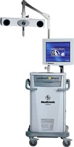
The innovative solution for FESS Functional Endoscopic Sinus Surgery (Technology Overview)
The Land marX® Element IGS System is the innovative solution for FESS, making image guidance simpler and more affordable. This fast, user-friendly system can help you quickly integrate IGS with your OR. Setup and registration are nearly automatic through auto-recognition of instruments and auto-progression of tasks.
The Element provides the tools you need to navigate within minutes. The system includes a straight probe, Ferguson 9FR suction and single-calibration curved suction. Used with the Element software, the curved suction is simply brilliant. After one calibration, you can navigate toward both maxillary sinuses and the frontal recess.
Call ENT & Allergy Specialists – Ear Nose and Throat Physicians and Surgeons at (859) 781-4900 for more information or to schedule an appointment.